

When Wilhelm Conrad Röntgen first published his very recent discovery of the X-ray in December of 1895 in an article entitled “On a New Kind of Rays, a Preliminary Communication,” he wrote about a new form of transparency in which “bodies behave to the X-rays as turbid media to light.”1 The invisible rays are described as a “medium” that penetrates objects and is revealed on screens. A floating technical surface acts as the most intimate witness of the otherwise hidden interior. An architecture is established that inverts the classical relationship between inside and outside, an architecture we still live in today, with our countless screens monitoring endless invisible flows. Architects, historians, and theorists quickly absorbed the new paradigm—developing an entire logic of the invisible in the early decades of the twentieth century that remains largely in place. New medical screens are today creating new forms of architecture as the relationship between inside and outside passes through another twist. New forms of intimacy are emerging.
Even before he mentions X-rays, Röntgen describes in the second paragraph of his paper a new concept of transparency closely linked to the idea of a “screen.” The screen is actually made of a piece of paper coated with a thin layer of “barium platinum cyanide” that glows fluorescent when exposed to the rays. He marvels at the fact that paper itself is very transparent when seen through such a “fluorescent screen,” so the screen is really just the thin layer of barium platinum cyanide. But it is not just a sheet of paper that is transparent. Even a thousand-page book placed behind the screen becomes transparent. “Thick blocks of wood are still transparent.” Tinfoil needs many layers to hardly cast a “shadow” on the screen and it takes a very thick sheet of aluminum to reduce the fluorescence.
1. The New Transparency
Röntgen’s screen showed, in the words of his first report, that “all bodies possess this same transparency, but in very varying degrees.”2 Transparency, therefore, is a property of seemingly opaque bodies, including the human body. In other words, it is not an effect. The X-ray is not something done to an object. The object is already transparent and the X-rays allow us to see it. The whole world is now understood to be transparent.
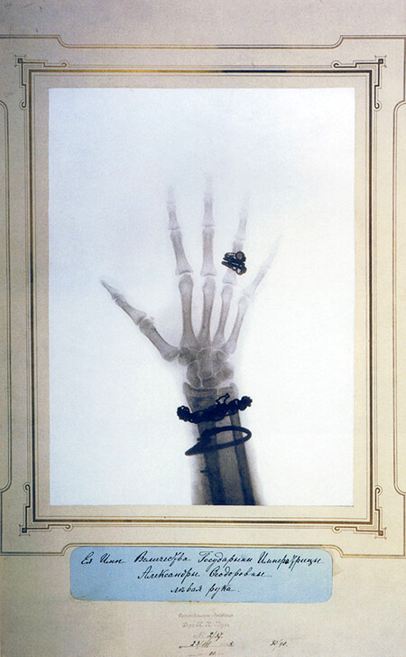
Having studied the transparency of many materials, including glass itself, which paradoxically is more opaque (because it contains lead), Röntgen looks through the human body: “If the hand be held before the fluorescent screen,” he writes, “the shadow shows the bones darkly, with only faint outlines of the surrounding tissues.”3 The famous X-ray image of the hand of his wife Bertha Röntgen, with her wedding ring on the third finger, taken only five days before he submitted the article for publication, is used as an illustration—as proof of the astonishing revelation.4 The image was crucial to the popular success of the invention.
Afraid of revealing his stunning discovery to his colleagues, Röntgen brought his wife to the laboratory the evening of December 22, 1895, after months of experimentation with inanimate objects, and exposed her hand to the X-rays for fifteen minutes, producing the first X-ray image of a human body. Upon seeing the image of her hand, Bertha Röntgen famously said, “I have seen my death,” anticipating a common popular reaction to such images.5
Röntgen’s article stimulated an enormous double reaction. Scientists all around the world seized on the idea and tried to replicate the experiment. The popular press ignited intense speculation about the possible uses and meaning of these images. Newspapers were galvanized by the idea of an invisible world. The image of Bertha’s hand gave way to a whole genre of such images in both scientific and popular publications. Countless X-ray images of hands were made by Röntgen and others soon after the article was published. Röntgen photographed the hand of Professor Albert von Kolliker, a famous anatomist and president of the Wurzburg Physical and Medical Society, during Röntgen’s first public lecture about the discovery, delivered on January 13, 1896. Summoned to Berlin by the emperor to report on the discovery, he photographed the hands of Wilhelm II and Empress Augusta Victoria. Later he also X-rayed the hands of the duke and duchess of York and the emperor and empress of Russia, among many other notables. The hand X-ray had become a new kind of intimate portrait and the icon of a new worldview, a worldview in which everything, no matter how seemingly impervious, becomes intimate.


Röntgen had named the mysterious phenomenon “X-rays” because he didn’t know what they were. Kolliker had proposed, after Röntgen’s Wurzburg lecture, that the new rays be called “Röntgen rays,” but self-effacing Röntgen preferred to continue to call them X-rays. The justification of the term “rays,” he claimed in that first article, lay in the “shadow pictures” produced by interposing a body between the source of the X-rays and a photographic plate or a screen. Röntgen wrote that in the two months before he photographed his wife’s hand, he had “observed and photographed many such shadow pictures,” including a set of weights inside a wooden box and a compass card and needle completely enclosed in a metal case.6
Röntgen considered this ability to make photographs of the “shadow pictures” on the screen of “special interest,” because it made it possible to “exhibit the phenomena so as to exclude the danger of error.”7 The main advantage for him, therefore, was to provide proof, to confirm the observations already made multiple times with the fluorescent screen. Soon after its initial publication, he sent reprints of the article together with prints of the X-ray images he had taken to many scientists, including Emil Warburg in Berlin and Henri Poincare in Paris.8 Warburg immediately added the X-ray images to an exhibition that was already mounted on the occasion of the anniversary of the Berlin Physical Society at Berlin University. This was the first public exhibition of X-ray images.
Without these images, the discovery of the X-rays would have been of less interest both in scientific and lay circles. It was front-page news in many newspapers worldwide. On January 5, 1886, Die Presse in Vienna was the first to report on the discovery, with an article entitled “Eine Sensationelle Entdeckung” (A Sensational Discovery).9 In their haste, they misspelled Röntgen’s name as “Routgen.” The next day, the London Standard cabled the following news release to the world, repeating the misspelled name:
The noise of war’s alarm should not distract attention from the marvellous triumph of Science which is reported from Vienna. It is announced that Professor Routgen of Würzburg has discovered a light which, for the purpose of photography, will penetrate wood, flesh and most other organic substances. The Professor has succeeded in photographing metal weights which were in a closed wooden case, also a man’s hand, which shows only the bones, the flesh being invisible.
When the London Standard reported on the story in the paper on January 7, 1896, they felt obliged to add: “The Presse assures its readers that there is no joke or humbug in the matter. It is a serious discovery by a serious German Professor.” The Frankfurter Zeitung published the news, also on January 7, and was the first newspaper to print the images. Newspapers around the world, from Paris Matin, to the Krakow-based Czas, to the New York Times, St. Louis Dispatch, and Sydney Telegraph, among many others, continued to sensationalize the discovery and anticipate its medical uses, something that Röntgen was skeptical about. Since only text could be sent by cable telegraphy, most of these early overseas reports were not illustrated—prompting skepticism among readers and journalists. Czas, for example, wrote: “the problem, although it seems an All Fools’ Day joke, is seriously considered in serious circles.”10
2. The Magic Screen
Many people followed on Röntgen’s invention, developing techniques for photographing the X-ray effect. Röntgen had refused several offers to patent his discovery, declaring that it belonged to humanity. He died in near poverty. From the beginning, he provided detailed accounts of his method, allowing others to experiment. Within a month of Röntgen’s publication, Josef Maria Eder (director of an Austrian institute for graphic processes and author of an early history of photography11) and the photo chemist Eduard Valenta published Versuche über Photographie mittelst der Röntgen’schen Strahlen (Research on Photography with Röntgen Rays), an album of fifteen photogravures made from X-rays, in which they described in great detail the procedure used and the improvements they had made to Röntgen’s apparatus.12 Human hands and feet, fish, frogs, a snake, a chameleon, a lizard, a rat, and a newborn rabbit were among the images in the album, a kind of zoo echoing the nineteenth-century naturalist albums of animals, insects, and plants, but also anticipating the new vision of photography of Moholy-Nagy and others in the 1920s. This new vision of photography would also become encyclopedic, as if the whole world had to be seen again, or more precisely, as if it were a whole new world. Moholy-Nagy would later write:
The passion for transparencies is one of the most spectacular features of our time. In x-ray photos, structure becomes transparency and transparency manifests structure. The x-ray pictures, to which the futurist has consistently referred, are among the outstanding space-time renderings on the static plane. They give simultaneously the inside and outside, the view of an opaque solid, its outline, but also its inner structure.13
What interests me here is precisely how X-ray images transformed the visual field long before the so-called avant-garde. The X-ray was a new kind of realism, which was in no way in opposition to a new kind of mysticism or spiritualism. William Crookes, the scientist who developed the cathode tube used by Röntgen, was the president of the Society for Psychical Research and within a year of the discovery of X-rays announced that they produced a new sense of reality based not on outer surfaces but inner vibrations, closer to consciousness itself, as Linda Henderson has pointed out.14 The X-ray was an optical and philosophical revolution that swept the world at astonishing speed. The first surgery with X-rays was carried out in the US within two months of the discovery, and the first fully fledged department of radiology was established at the Glasgow Royal Infirmary within a year.
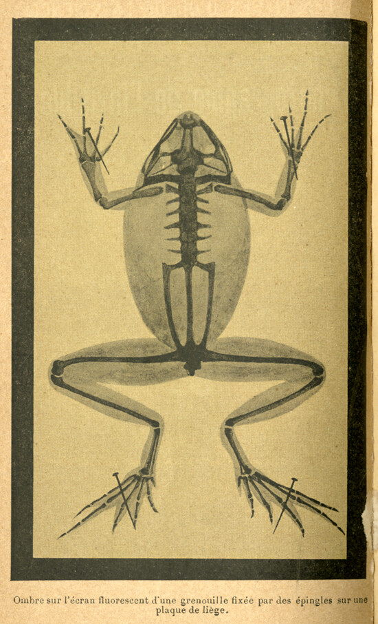

It is important to note that the albums of X-rays that proliferated everywhere were still presented as images of screens. Even books aimed at popularizing science, such as the 1898 French book La Photographie de l’Invisible, carefully note below each image that it is a “shadow on a screen.” For example, under the X-ray of a frog we read: “ombre sur l’écran fluorescent d’une grenouille fixée par des épingles sur une plaque de liège” or “ombre d’une main sur un écran au platinocyanure de barium” (shadow of a hand on a screen of platinum cyanure of barium).15 The caption is needed because the screen itself disappears. It has the same color as the page in the publication. The caption reminds the reader that there is a screen there, a screen that was originally paper. The “shadow image” takes the place and the modality of a drawing, a ghostly trace hanging before the viewer and offering a deep gaze into the secrets of a body or even of the cosmos itself. The floating, disappearing screen becomes the most powerful of instruments.
Röntgen was fascinated that he could also produce the effect directly on a photographic plate. Within a year of the discovery of X-rays, Eastman developed a special plate for X-rays; a thin transparent surface would eventually take over the responsibility of the screen when Eastman introduced film, replacing the glass photographic plate. But the doubling, the eerie status of the shadow image, remained. The photograph of Bertha’s hand was the image of an image, the proof of what Röntgen had seen countless times on the screen. It is still the image of the screen.
This magical and threatening screen effect rippled through society, becoming a new form of spectacle. It was as if nothing could be seen the same way. Everything needed to be rethought. Every field seemed to be affected by the magic screen—science and medicine of course, but also policing and entertainment, religion and spiritualism (where many seized upon the X-ray as proof of what they believed all along). The screen was a site of intense speculation.
From the very beginning this visual revolution was understood as an assault on privacy, and even as a form of indecency. The London newspaper Pall Mall Gazette wrote in 1896: “We are sick of the Röntgen rays … It is now said that you can see other people’s bones with the naked eye … On the revolting indecency of this there is no need to dwell.”16 Cartoons and comical poems explored the new space of exposure. The fear that X-rays would allow people to see through clothing existed from the beginning. A poem in Electrical Review in 1896, for example, goes:
The Roentgen Rays, the Roentgen Rays,
What is this craze?
The town’s ablaze
With the new phase
Of X-ray’s ways.
I’m full of daze,
Shock and amaze;
For nowadays
I hear they’ll gaze
Thro’ cloak and gown—and even stays,
These naughty, naughty Roentgen Rays.17
Shortly after the invention, merchants offered X-ray–proof underwear (as happened again in recent years when customs and security introduced full-body scanners in airports in 2007). And a New Jersey assemblyman is supposed to have introduced a bill to ban X-ray opera glasses, should they ever be invented. Thomas Edison, who exhibited X-rays to the public in the New York Electrical exhibition of 1896, even imagined that the X-ray would eventually read people’s thoughts.
The X-ray was an immense form of entertainment. There were X-ray machines in every fair, scientific and popular. A leaflet distributed at an 1896 exhibition at the Crystal Palace, London reads: “Before Leaving the Exhibition, ‘SEE’ the Wondrous X-rays, the greatest scientific Discovery of the Age … X-ray Photographs Taken.” In Paris, the Grands Magazines Dufayel alternated demonstrations of an X-ray machine with demonstrations of the Lumière brothers’ moving pictures. Customers could have an X-ray taken of their hand or their feet as a souvenir. In fact, cinematography and X-rays were discovered within a few months of each other, in late 1895. X-ray equipment was bought not just by scientist but by entrepreneurs, some of whom believed that X-rays would offer more entertainment value than cinema. Business trade journals carried ads from impresarios trying to exchange their movie projectors for X-ray equipment.18 In 1896, Bloomingdales hired Columbia University physics senior Herbert Hawks to conduct public demonstrations of X-rays. There were X-ray studios in all major cities. X-ray slot machines were installed in Chicago; you could have an X-ray for $1.

3. Building in the New Visual Field
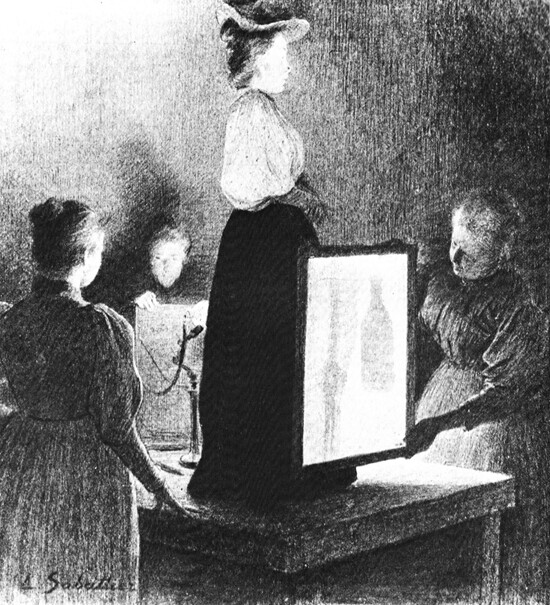
X-rays were almost immediately used for policing in customs checkpoints, where suitcases and people were subjected to exposure. In Paris railways stations, the police subjected passengers and their luggage to X-rays as early as 1898. An illustration in a Parisian newspaper shows how a woman hiding a bottle of liquor under her dress is exposed by the machine as the glass with lead becomes visible next to her femur in the X-ray.
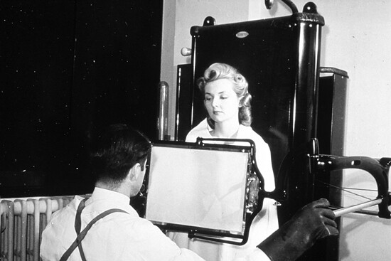
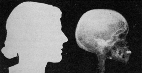
What is crucial here is the architecture of the scene. She is suspended behind a floating screen, held in place by an assistant. She is occupying a new space of radical exposure. The X-ray was architectural from the beginning and remains so, as can be seen in image after image, like the photograph of an attractive blond woman behind a screen made in 1940 as a publicity image to reassure the public that radiation from X-rays was negligible. She is occupying a new technological space defined by a screen rather than walls, a glowing screen with its shadow image. We are still in the territory of Bertha Röntgen and the ur-image of her hand. The mysteries of the interior are brought to the surface by a screen and the flesh becomes just a faint outline. The body is literally turned inside out.

Western architecture, at least since the Italian Renaissance, has modeled itself on the human body. With the arrival of X-rays, the body is inverted—the inside becomes the outside. Modern architecture absorbed the logic of the screen and even of the shadow image. In glass architecture the logic of the X-ray applies. There is an outer screen that disappears in order to register a ghostly image of the inside. It is X-ray architecture. As with Röntgen’s transformative images, X-ray architecture becomes an image of an image—the effect of an X-ray, rather than an actual X-ray. It’s not so much that the inside of the building is exposed but that the building represents exposure and this exposure occurs on a screen. Glass is called on to simulate transparency.
This X-ray effect was integral to a new discourse about transparency. Arthur Korn’s remarkable 1929 book Glas im Bau und als Gebrauchsgegenstand (Glass in Construction and as a Commodity), for example, catalogs the new use of glass in architecture and remarks, as if surprised, that “the outside wall is no longer the first impression one gets of a building. It is the interior, the spaces in depth and the structural frame which delineates them, that one begins to notice through the glass wall … Glass is noticeable yet not quite visible. It is the great membrane, full of mystery, delicate yet tough.”19 This sense of mystery, which X-rays share, infuses Korn’s book, as in photographs of the Bauhaus building in Dessau where the glass wall is a kind of ripple—the volume of the building within looms without definition.

Korn’s discussion of transparency is an uncanny echo of Röntgen’s discussion of new forms of “transparency” in the very first publication of his discovery of the X-ray. Just as the body of the Bauhaus building appears strangely blurry through the not-quite-visible glass, Röntgen writes about the flesh becoming a kind of mysterious shadow while the bones are perfectly visible.
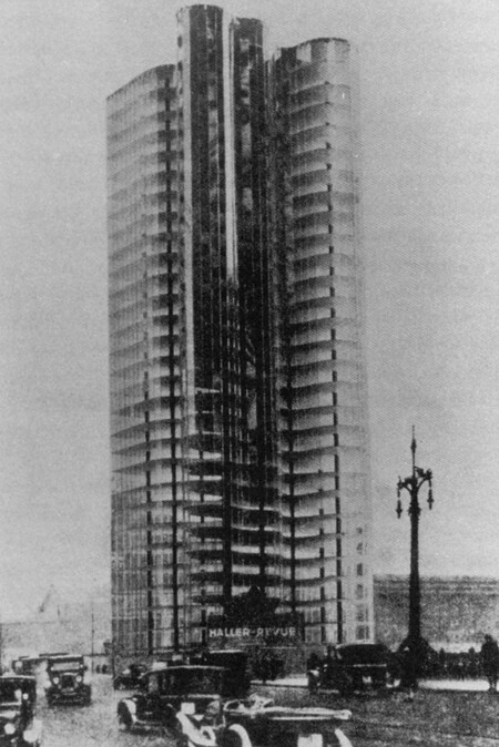
Modern buildings even started to look like medical images. The impact of the technology of the X-ray, the dominant diagnostic tool for lung tuberculosis, is evident in the work of many avant-garde architects of the early decades of the twentieth century. Mies van der Rohe wrote about his work as “skin and bones” architecture, and rendered his Friedrichstrasse Skyscraper of 1919 and his Glass Skyscraper of 1922 as if seen through an X-ray machine. Mies was deeply interested in X-ray images and used them as illustrations in his articles, as in the April 1926 issue of G. He even put an image of a bone alongside his glass skyscraper in Merz to drive the point home.

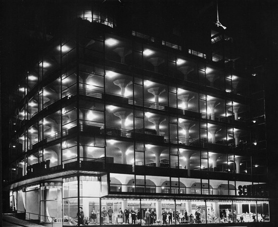
Mies was not alone. Books on modern architecture are filled with images of glowing glass skins revealing inner bones and organs; they look like albums of X-rays, reminiscent of the X-ray atlases that proliferated in the early decades of the twentieth century.20 Think, for example, about Le Corbusier’s project for the Glass Skyscraper (1925), Walter Gropius’s Bauhaus (1925–26), Brinkman & Van der Vlugt’s Van Nelle Factory (1925–27) in Rotterdam, Mendelsohn’s Schocken Department Store (1926–28) in Stuttgart, George Keck’s Crystal House (1933–34) at the World’s Columbian Exhibition in Chicago, Paul Nelson’s Suspended House (1935), Frits Peutz’s Schunck Glass Palace (1935) in Heerlen, and countless other examples. This is more than a dominant aesthetic. It is a symptom of a deep-seated philosophy of design deriving from medical discourse.
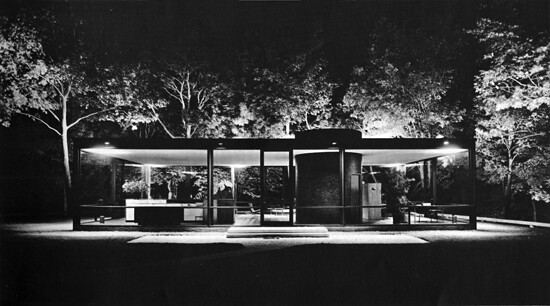
The development of the X-ray and that of modern architecture coincide; they evolved in parallel. If experiments with glass were numerous in the early years of the twentieth century, they still tended to be isolated esoteric projects by avant-garde architects—many developed as temporary buildings for fairs. Only by the mid–twentieth century did the see-through house become realized in Mies’s Farnsworth House (1945–51) in Plano, Illinois, and Philip Johnson’s Glass House (1949) in New Canaan, Connecticut. Just as the X-ray exposes the inside of the body to the public eye, modern architecture exposes its interior. This exposure becomes a mass phenomenon with the picture window at mid-century at exactly the same time that the X-ray itself is becoming a mass phenomenon.

4. A Glass House Should Hold No Terrors
It was not just the house that had to be see-through. Everything from Pyrex cookware, to Saran Wrap, to windows in ovens and washing machines exposed their contents. Likewise, everything was subject to X-rays—even cars, as in a 1946 image of a Jeep featured in Life (“World’s Biggest X-Ray”) and used in the exhibition “Parallel of Life and Art” at the Institute of Contemporary Arts in London in 1953. The front cover of the catalog has a 1941 X-ray image of a man using an electric shaver, taken from László Moholy-Nagy’s 1947 book Vision in Motion, where it is described as the work of two doctors in a New Jersey laboratory. The image had been published in Mechanix Illustrated, which is probably where Moholy-Nagy got it. But Moholy-Nagy also picked up images of X-rays from a 1923 issue of Wendingen magazine—which goes to show that architectural magazines were publishing X-ray images from early on.
But by the time of Moholy-Nagy’s book, X-rays had evolved from being radical images representing the hidden truth of things to becoming almost routine elements of everyday life. Starting in the 1930s, shoe stores used X-ray machines for shoe fittings without any kind of protection from radioactivity, which wasn’t banned until the 1970s. Also in the 1930s the mass X-raying of citizens on a regular basis started. With this development, the now-visible interior of the body became not just a tool for diagnosis but also the site of a new form of public surveillance. The postwar mobilization against TB included programs for the mass X-ray surveying of the entire population using mobile X-ray machines in places such as department stores, workplaces, schools, suburban streets, and public markets. Over a period of half a century, an experimental medical tool had been transformed into a mechanism of surveillance for the whole population.
The association between X-rays and glass houses became commonplace in mid-century popular culture. For example, in Highlights and Shadows, a 1937 Kodak Research Laboratories film on the virtues of X-rays for disease prevention by the filmmaker-radiographer James Sibley Watson, Jr., a woman wearing a swimsuit is shown strapped to a laboratory table while her body is subjected to X-rays. As her photographic image gives way to the image of her X-rayed body, the narrator declares: “This young lady, to whom henceforth a glass house should hold no terrors, will after an examination of her radiographs, be reassured that she is indeed physically fit.”21 The glass house acted as a symbol of both the new form of surveillance and health.
A similar set of associations can be found in the discourse surrounding canonic works of modern architecture. In an interview in House Beautiful, Edith Farnsworth, a successful doctor in Chicago, compared her famous weekend house, designed by Mies in 1949, to an X-ray:
I don’t keep a garbage can under my sink. Do you know why? Because you can see the whole “kitchen” from the road on the way in here and the can would spoil the appearance of the whole house. So I hide it in the closet further down from the sink. Mies talks about “free space”: but his space is very fixed. I can’t even put a clothes hanger in my house without considering how it affects everything from the outside. Any arrangement of furniture becomes a major problem, because the house is transparent, like an X-ray.22
The use of the metaphor of the X-ray was not accidental. It is not by chance that Farnsworth goes on to say of her house: “There is already the local rumor that it’s a tuberculosis sanatorium.”23 Modern architecture was literally presented and understood as a piece of medical equipment.

Modern architecture cannot be understood outside tuberculosis and its dominant diagnostic tool, the X-ray. Indeed, the principles of modern architecture seem to have been taken straight out of a medical text on the disease. A year before the German microbiologist Robert Koch discovered the tubercle bacillus in 1882, a standard medical book gave as the causes of the disease “unfavorable climate, sedentary indoor life, defective ventilation and deficiency of light.”24 It took a long time for these notions to lose credibility, as Susan Sontag writes: “The TB patient was thought to be helped, even cured, by a change in environment. There was a notion that TB was a wet disease, a disease of humid and dank cities. The inside of the body became damp (‘moisture in the lungs’ was a favored locution) and had to be dried out.”25
Modern architects offered health by providing exactly such a change of environment. Nineteenth-century architecture was demonized as unhealthy, and sun, light, ventilation, exercise, roof terraces, hygiene, and whiteness were offered as means to prevent, if not cure, tuberculosis. The publicity campaign of modern architecture was organized around contemporary beliefs about tuberculosis and fears of the disease—being deeply affected by the primary diagnostic tool of the chest X-ray. Modern architecture not only thinks of itself as providing sanatorium conditions for everyday life but even thinks of buildings as diagnostic instruments with the power of an X-ray. As Le Corbusier put it in L’art decoratif d’aujourd’hui in 1925: “If the house is all white … everything stands out from it and is recorded absolutely, black on white; it is honest and dependable … It is rather like an X-ray of beauty.”26
Le Corbusier’s metaphor was no accident. Diagnosis of tuberculosis continued to be difficult; physicians often confused it with other illnesses, including bronchitis, chronic indigestion, malaria, neurasthenia, and typhoid fever. To evaluate the condition, they needed to see inside the body. X-ray technology had been available in sanatoriums since the beginning of the century, and by the 1920s, the X-ray was a routine part of the examination of those with visible symptoms. Screening the body for tuberculosis meant optically penetrating areas of the body previously invisible. X-rays created a new kind of vision, a new paradigm of truth that architects could not resist. Nothing could have been more modern.
“The TB sufferer is a wanderer” wrote Susan Sontag in one her notebooks in her archives in UCLA. The patient travels to find a cure. “There is a geography of health.”27 This nomadic figure is also the paradigmatic client of modern architecture that enters modern buildings like entering any other medical apparatus. Architecture here is less about shelter and more about a kind of exposure—X-ray exposure. There is a whole architecture of health organized around a new kind of image. The discourse about transparency in modern architecture is but an echo of the discourse about transparency that was already part of Röntgen’s first scientific paper announcing the discovery of X-rays in 1895 and immediately captivating the popular imagination: the idea that one could see through buildings and clothing challenged all assumptions and social protocols about privacy and psychological well-being.
As X-rays became indelibly associated with tuberculosis, the tuberculosis patient became the paradigm of this new way of thinking about bodies, objects, and psychologies. “TB makes the body transparent” writes Sontag in another note in her archives, “while to have cancer is to become more than normally opaque,” perhaps because cancer turns the very structure of the body into a blur. “The X-ray permits one, often for the first time, to see one’s inside, to become transparent to oneself,” she writes.28 Indeed, the tuberculosis patients in the sanatorium of Thomas Mann’s The Magic Mountain carry their X-rays, or those of their loved ones, in their breast pockets. When Clavdia Chauchat, Hans Castor’s love object, leaves the sanatorium, she gives him her X-ray as a memento:
Then he flung himself into his chair, and drew out his keepsake, his treasure, that consisted, this time, not of a few reddish-brown shavings, but a thin glass plate, which must be held toward the light to see anything on it. It was Clavdia’s x-ray portrait, showing not her face, but the delicate bony structure of the upper half of her body, and the organs of the thoracic cavity, surrounded by the pale, ghostlike envelope of flesh. How often had he looked at it, how often pressed it to his lips in the time which since then had passed and brought its changes with it—such changes as, for instance, getting used to life up here without Clavdia Chauchat, getting used, that is, to her remoteness in space!29
The X-ray is a kind of self-exposure, a new, more intimate kind of portrait. The intrusive logic of medical and police surveillance, with the body unable to resist a newly penetrating gaze, gives way to a tender intimacy. The attempt to discipline the body—with a new regime of synchronized medical, technological, and architectural protocols—produces new psychological, social, philosophical, and emotional interactions. The seemingly fragile cloudy space of the X-ray becomes an architecture in its own right that can be inhabited, and is inhabited. All the ostensible sharpness and clarity of modern architecture gives way to soft layers of reflections and translucencies. X-ray architecture is an occupiable blur.
Wilhelm Röntgen, “On a New Kind of Rays,” Nature, January 23, 1896, 274–76; English translation of the 1895 original text “Uber eine neue art von strahlen,” published in the Sitzungsberichte der Physikalisch-Medizinischen Gesellschaft in Würzburg 137, December 28, 1895, 132–41.
Ibid.
Ibid.
The original article did not include illustrations, but the English version published in Nature a few weeks later did.
Multiple sources, including Röntgen Centennial: X-rays in Natural and Life Sciences, ed. Gottfried Landwehrand and A. Hasse (Singapore: World Scientific, 1997), 7–8.
Röntgen, “On a New Kind of Rays.”
Ibid.
Röntgen also sent the reprint and images to the following scientists: Arthur Schuster in Manchester, Friedrich Kohlrauch in Gottingen, Lord Kelvin in Glasgow, and Franz Exner in Vienna.
It was Franz Exner who alerted the Vienna newspaper to Röntgen’s discovery.
A. Urbanik, “History of Polish Gastrointestinal Radiology,” Journal of Physiology and Pharmacology, vol. 54, no. 3 (2003): 211.
Josef Maria Eder, Ausfuhrliches Handbuch Der Photographie (Halle, 1884).
Josef Maria Eder and Eduard Valenta, Versuche über Photographie mittelst der Röntgen’schen Strahlen (Vienna: R. Lechner & Halle, Wilhelm Knapp, 1896).
László Moholy-Nagy, “Space-Time Problems,” first published in American Abstract Artists Yearbook (New York, 1946). Reprinted in Vision in Motion (Chicago, 1947), 252.
Linda Dalrymple Henderson, Duchamp in Context: Science and Technology in the Large Glass and Related Works (Princeton: Princeton University Press, 1998). See also Tom Gunning, “Invisible Worlds, Visible Media,” in Brought to Light: Photography and the Invisible 1840–1900, ed. Corey Keller (New Haven, CT: Yale University Press).
L. Aubert, La Photographie de l’Invisible: les rayons X suivi d’un glossaire (Paris: Les livres d’or de la science, 1898).
Pall Mall Gazette, March 1896. Quoted in Jon Queijo, Breakthrough!: How the 10 Greatest Discoveries in Medicine Saved Millions and Changed Our View of the World (London: FT Press, 2010).
Wilhelma, Electrical Review, April 17, 1896.
Tom Gunning, “Invisible Worlds,” 52.
Arthur Korn, introduction to the first edition of Glas im Bau (1929); quote from the introduction of the English edition by Barrie & Rockliff.
Rudolf Grashey’s Typische Röntgenbilder von normalen Menschen, for example, went through six printings between 1905 and 1939. See Lorraine Daston and Peter Galison, “The Image of Objectivity,” in “Seeing Science,” special issue, Representations 40 (Autumn 1992): 81–128.
James Sibley Watson, Jr., Highlights and Shadows (Kodak, 1937), quoted in Lisa Cartwright, Screening the Body: Tracing Medicine’s Visual Culture (Minneapolis: University of Minnesota Press, 1995), 155.
Edith Farnsworth quoted in Joseph A. Barry, “Report on the American Battle between Good and Bad Modern,” House Beautiful, May 1953, 270.
Edith Farnsworth, “Memoirs” (unpublished manuscript), quoted in Alice T. Friedman, Women and the Making of the Modern House: A Social and Architectural History (New York: Abrams, 1998), 143.
August Flint and William H. Welch, The Principles and Practice of Medicine (5th edition, 1881), cited in Susan Sontag, Illness as Metaphor (New York: Vintage Books, 1979), 53.
Ibid., 14–15.
Le Corbusier, L’art decoratif d’aujourd’hui (Paris: G. Crès et Cie, 1925),190; my translation.
Susan Sontag Archives, UCLA, Box 43.
Ibid.
Thomas Mann, The Magic Mountain (New York: Alfred Knopf, 1945), 348–49.